
The Jeil Medical Orthodontic Anchor Plate System: A Comprehensive Guide
Orthodontic treatment often requires stable anchorage to achieve desired tooth movements. While mini-implants have become a popular tool, the Orthodontic Anchor Plate System by Jeil Medical offers a distinct and highly versatile alternative for challenging cases. This system, utilizing T and Y plates, provides robust skeletal anchorage, expanding the possibilities for complex orthodontic mechanics.
Understanding T and Y Plates: Versatility in Action
The Jeil Medical system primarily features two types of anchor plates: T-plates and Y-plates. Their unique designs cater to different anatomical locations and biomechanical requirements:
· T-Plates: These plates, shaped like the letter 'T', are typically used in areas offering broad bone contact, such as the retromolar pad region or the palate. Their wider base provides excellent stability, making them ideal for en masse retraction, molar protraction, or distalization where significant force application is needed. They are particularly effective when a broad, flat surface for anchorage is available.
· Y-Plates: Designed with a 'Y' shape, these plates are often preferred in areas with more complex anatomy or where a more distributed force application is desired, such as the anterior maxilla or mandible. The bifurcated arms of the Y-plate allow for placement around anatomical structures and can accommodate different vectors of force, providing greater flexibility in anchorage planning. They are especially useful for intrusion of anterior teeth or addressing asymmetries.
Differentiating from Mini-Implants: A Matter of Stability and Force
While both anchor plates and mini-implants provide skeletal anchorage, they differ significantly in their application and capabilities:
· Stability and Load Bearing: Anchor plates offer superior stability and can withstand much greater orthodontic forces compared to mini-implants. This is due to their larger surface area of bone contact and fixation with multiple screws. Mini-implants, being smaller and fixed with a single point of contact, are more prone to mobility under heavy loads.
· Indications: Mini-implants are excellent for individual tooth movements or mild to moderate anchorage needs. Anchor plates, on the other hand, are invaluable for complex movements requiring absolute anchorage, such as en masse distalization of entire arches, severe open bites requiring significant intrusion, or cases with compromised periodontal support where traditional anchorage is insufficient.
· Placement and Removal: Mini-implant placement is a simpler chairside procedure, often performed by the orthodontist. Anchor plate placement is a minor surgical procedure, requiring the expertise of an oral and maxillofacial surgeon. Similarly, removal of anchor plates, while generally straightforward, also requires a surgical approach.
Advantages and Disadvantages of Anchor Plates
Advantages:
· Absolute Anchorage: Provides unyielding stability for the most challenging orthodontic movements.
· Heavy Force Application: Can withstand significant orthodontic forces, enabling rapid and efficient tooth movement.
· Versatility: T and Y plates, along with various sizes, allow for adaptation to diverse anatomical locations and treatment goals.
· Predictable Outcomes: Reduces unwanted side effects and improves the predictability of treatment results.
· Eliminates Patient Compliance Issues: Unlike headgear or elastics, anchor plates are fixed, eliminating the need for patient cooperation.
Disadvantages:
· Surgical Procedure: Requires a minor surgical procedure for placement and removal.
· Cost: Generally more expensive than mini-implants.
· Patient Discomfort: Post-operative discomfort and swelling are possible, though usually manageable.
· Healing Time: Requires a short healing period before orthodontic forces can be applied.
· Visibility: In some locations, the plate may be palpable or visible, though rarely aesthetically significant.
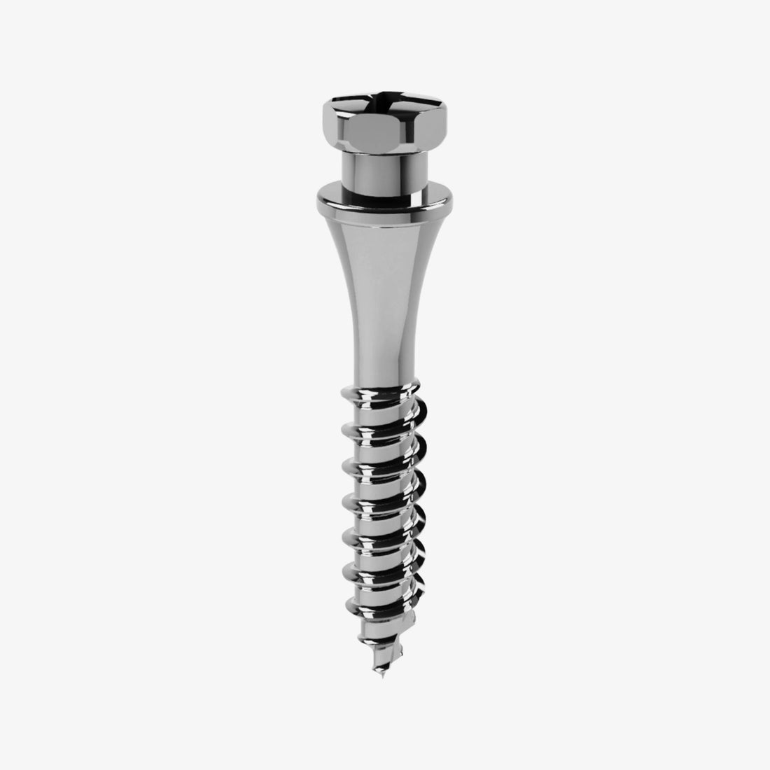
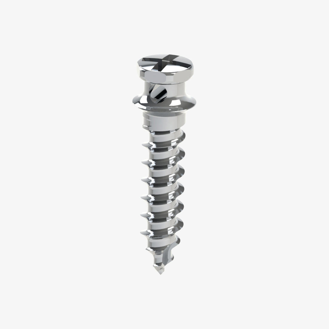
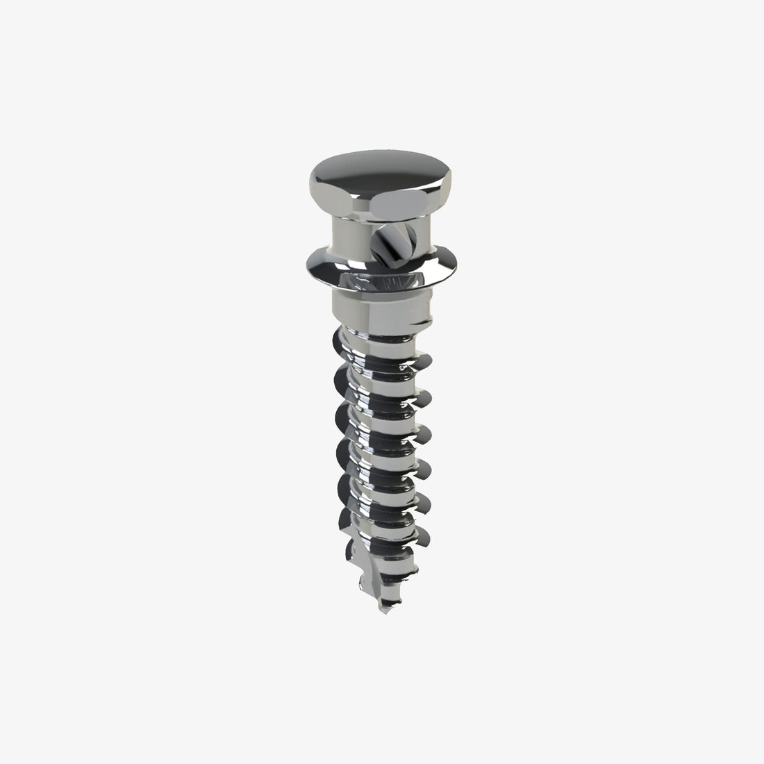
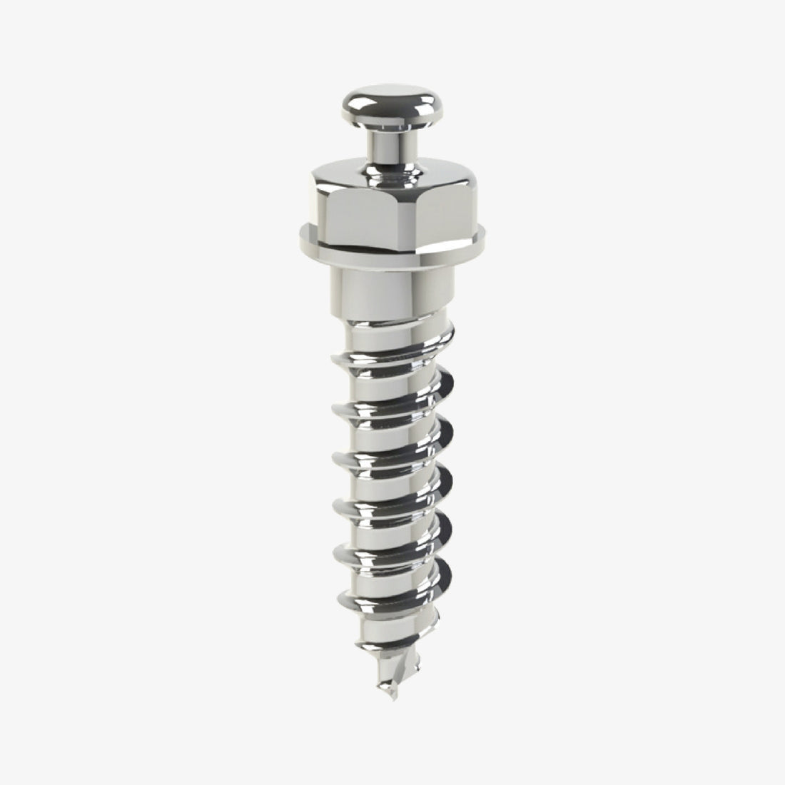
Fixation Screws: The Role of 2.0 Auto screws
The Jeil Medical Orthodontic Anchor Plates are securely fixed to the bone using 2.0 auto screws. These self-drilling and self-tapping screws are specifically designed for optimal bone engagement and stability.
Main Characteristics and Advantages of 2.0 Auto screws:
· Self-Drilling and Self-Tapping: Eliminates the need for pre-drilling a pilot hole in many cases, simplifying the surgical procedure and reducing chair time. This feature also minimizes heat generation during insertion.
· Optimal Thread Design: The unique thread design ensures excellent primary stability and strong bone grip, crucial for resisting orthodontic forces.
· Biocompatible Material: Typically made from medical-grade titanium alloy, ensuring excellent biocompatibility and minimizing the risk of adverse reactions.
· Strength and Durability: Designed to withstand the forces encountered during insertion and throughout the orthodontic treatment period.
· Ease of Insertion and Removal: While providing strong fixation, their design allows for relatively straightforward insertion and removal by a skilled surgeon.
The Indispensable Orthodontist - Surgeon Collaboration
The successful application and utilization of the Orthodontic Anchor Plate System critically depend on a seamless collaboration between the orthodontist and the oral and maxillofacial surgeon.
· Orthodontist's Role: The orthodontist diagnoses the case, formulates the treatment plan, and identifies the specific anchorage needs. They determine the ideal location, type (T or Y), and size of the anchor plate required to achieve the desired tooth movements. They also plan the biomechanics and force application post-placement.
· Surgeon's Role: The surgeon is responsible for the precise and sterile surgical placement of the anchor plate. They ensure proper anatomical positioning, secure fixation, and minimize surgical trauma. They also manage post-operative care and, when necessary, perform the removal of the plate.
Effective communication and joint planning between these two specialists are paramount. This ensures that the plate is placed in a position that is both surgically safe and orthodontically effective, maximizing the chances of successful treatment outcomes.
Detailed Step-by-Step Plan for Anchor Plate Application
The following outlines the general steps involved in the effective application of an orthodontic anchor plate in a patient's mouth. This is a surgical procedure and should only be performed by a qualified oral and maxillofacial surgeon in a sterile environment.
1. Pre-operative Assessment and Planning (Orthodontist & Surgeon):
l Thorough clinical examination and radiographic analysis (panoramic, cephalometric, CBCT scans if needed) to determine bone quality, quantity, and anatomical landmarks.
l Joint discussion and decision on the ideal plate type (T or Y), size, and precise placement location.
l Review of patient's medical history and current medications.
l Fabrication of a surgical guide if necessary for precise placement, especially in complex cases.
2. Patient Preparation:
l Informed consent obtained from the patient.
l Pre-operative instructions provided (e.g., fasting if general anesthesia is used, oral hygiene).
l Administration of prophylactic antibiotics as per protocol.
l Patient positioned comfortably in the dental chair.
3. Anesthesia:
Local anesthesia is typically administered to the surgical site. General anesthesia or conscious sedation may be used for anxious patients or complex cases.
4. Sterile Field Establishment:
l The surgical area is meticulously disinfected with an antiseptic solution (e.g., chlorhexidine).
l Sterile drapes are placed to maintain a sterile field.
5. Incision and Flap Reflection:
l A small, precise incision is made in the mucosa overlying the chosen placement site.
l A mucoperiosteal flap is carefully reflected to expose the underlying bone. The extent of the flap depends on the size of the plate and anatomical considerations.
6. Bone Preparation (if necessary):
l In some cases, minimal bone contouring or removal of sharp edges may be required to ensure intimate contact of the plate with the bone.
l The bone surface is meticulously cleaned.
7. Plate Adaptation and Positioning:
l The selected Jeil Medical anchor plate (T or Y) is carefully adapted to the bone surface to ensure passive fit and optimal contact.
l The surgical guide, if used, ensures precise orientation and position.
8. Screw Fixation:
l The 2.0 auto screws are used to fix the plate securely to the bone. Typically, 3 screws are used per plate.
l Screws are inserted slowly and under controlled pressure to prevent overheating of the bone.
l Care is taken to avoid vital anatomical structures (nerves, blood vessels, tooth roots).
9. Verification of Stability:
The surgeon gently tugs on the plate to confirm its absolute stability and lack of mobility.
10. Irrigation and Closure:
l The surgical site is thoroughly irrigated with sterile saline to remove any debris.
l The mucoperiosteal flap is repositioned and secured with sutures (resorbable or non-resorbable).
11. Post-operative Instructions and Care:
l Patient provided with clear instructions regarding pain management, oral hygiene, diet restrictions, and follow-up appointments.
l Prescription for pain medication and possibly antibiotics.
l Ice packs recommended for swelling management.
12. Healing Period:
A healing period of typically 2-4 weeks is recommended before orthodontic forces are applied to the anchor plate, allowing for initial osseointegration and soft tissue healing.
13. Orthodontic Force Application (Orthodontist):
Once the healing period is complete and the plate is stable, the orthodontist can begin applying the planned orthodontic forces to the anchor plate.
In conclusion, the Jeil Medical Orthodontic Anchor Plate System, with its versatile T and Y plates and secure 2.0 auto screw fixation, represents a powerful tool in the orthodontist's arsenal for managing complex cases. Its ability to provide absolute skeletal anchorage significantly expands treatment possibilities and enhances the predictability of outcomes, provided there is close and effective collaboration between the orthodontist and the oral and maxillofacial surgeon.


 0745 100 497


