Soft Tissue Management Around Skeletal Anchorage: Preventing Inflammation, Irritation and Hyperplasia
Skeletal anchorage has become an indispensable tool in modern orthodontics, enabling predictable and efficient tooth movements that were once challenging or impossible. While the stability and biomechanical advantages of mini-implants and anchor plates (such as those from Jeil Medical) are well-established, their success is not solely dependent on bone integration. The health and stability of the surrounding soft tissues play an equally crucial role in ensuring long-term success, preventing complications, and maintaining patient comfort.
Improper soft tissue management can lead to inflammation, irritation, pain, infection, and even hyperplasia (overgrowth of tissue), potentially compromising the anchorage device and overall treatment. This article explores key strategies for preventing these soft tissue complications around orthodontic mini-implants and anchor plate connections.
Understanding the Interface: Soft Tissue Challenges
The area where the mini-implant or anchor plate transgingival component exits the mucosa is a critical interface. Unlike dental implants, orthodontic mini-implants and anchor plates are not designed for osseointegration of the transmucosal part. They rely on a healthy, tight soft tissue seal to prevent bacterial invasion and maintain stability.
Common soft tissue issues include:
l Inflammation (Peri-implantitis/Pericoronitis): Redness, swelling, tenderness, and bleeding around the mini-implant or plate abutment, often due to plaque accumulation or mechanical irritation.
l Irritation: Discomfort caused by the mechanical friction of the appliance against the soft tissues, particularly if components are sharp, bulky, or placed improperly.
l Hyperplasia: Overgrowth of gingival tissue around the mini-implant or plate, often a response to chronic inflammation or irritation, making oral hygiene difficult and potentially leading to pocket formation.
l Infection: In severe cases, inflammation can progress to localized infection, requiring antibiotic treatment or even removal of the anchorage device.
Strategic Approaches to Soft Tissue Management
Effective soft tissue management involves a multi-faceted approach, encompassing careful site selection, meticulous surgical technique, appropriate post-operative care, and rigorous oral hygiene protocols.
1. Meticulous Site Selection and Planning
Before placement, comprehensive planning, often utilizing CBCT imaging, is paramount.
l Adequate Keratinized Gingiva: Whenever possible, mini-implants should be placed through keratinized gingiva rather than movable alveolar mucosa. Keratinized tissue is more resilient, less prone to inflammation, and easier for patients to clean. While not always feasible (e.g., in the retromolar area for some anchor plates), prioritizing attached gingiva where available significantly improves soft tissue health.
l Avoidance of Muscle Attachments: Placement near muscle attachments (e.g., buccal shelf, mentalis muscle area) can lead to constant soft tissue movement, increasing irritation and discomfort.
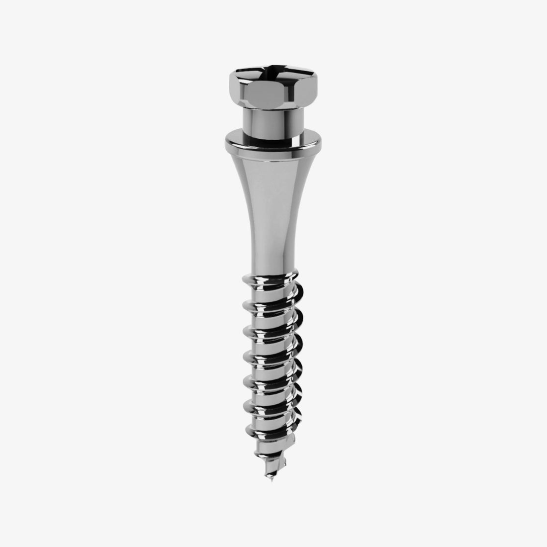
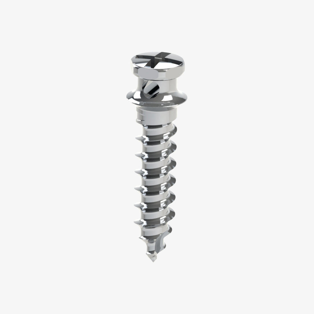
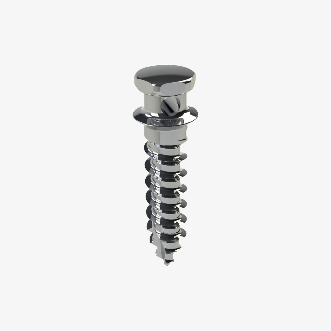
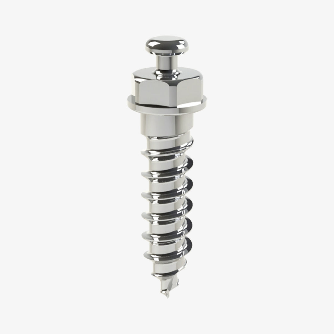
l Sufficient Space for Abutment: Ensure adequate space for the mini-implant head or the anchor plate connection component, preventing impingement on adjacent soft tissues or teeth. Jeil Medical offers various mini-implant head designs and anchor plate connections (e.g., button, hook) that require different clearance, necessitating careful pre-operative assessment.
l Access for Oral Hygiene: Choose a site that allows the patient easy access for cleaning, which directly impacts the ability to maintain a plaque-free environment.
2. Precise Surgical Technique
The surgical placement of both mini-implants and anchor plates (including those from Jeil Medical) directly impacts soft tissue integrity.
l Minimally Invasive Approach: For mini-implants, a non-flap technique is generally preferred to minimize tissue trauma. For anchor plates, while a flap is necessary, the incision should be designed to minimize tension upon closure.
l Controlled Drilling and Insertion: Overheating the bone during drilling or excessive torque during insertion can lead to localized tissue necrosis or inflammation. Using appropriate drills, irrigation, and controlled speed is crucial.
l Proper Emergence Profile: The mini-implant or plate component should emerge cleanly through the mucosa, avoiding sharp edges that could irritate the tissue. The design of Jeil Medical mini-implants aims for a smooth, transmucosal portion to facilitate this.
l Passive Fit of Attachments: Any attachments (e.g., orthodontic wires, springs, elastics) connected to the mini-implant or plate should have a passive fit and not impinge on the surrounding soft tissue. Adjustments should be made if any pressure points are observed.
3. Post-Operative Care and Instructions
Effective post-operative management is critical for initial healing and preventing complications.
l Analgesics and Anti-inflammatories: Prescribe appropriate pain medication and anti-inflammatory drugs (e.g., NSAIDs) to manage post-operative discomfort and reduce swelling.
l Antiseptic Mouth Rinses: Chlorhexidine gluconate (0.12%) mouth rinses twice daily for 7-10 days can significantly reduce bacterial load and promote initial soft tissue healing around the surgical site.
l Soft Diet: Advise patients to follow a soft diet initially to avoid mechanical trauma to the surgical area.
l Cold Compresses: Application of ice packs externally for the first 24-48 hours can help minimize swelling.
l Avoidance of Manipulation: Instruct patients to avoid touching or manipulating the mini-implant or plate with their tongue or fingers.
4. Rigorous Oral Hygiene Protocol
This is arguably the most crucial long-term strategy. Plaque accumulation is the primary cause of inflammation and hyperplasia.
l Gentle Brushing: Instruct patients to gently brush around the mini-implant or plate site with a soft toothbrush. Small, interdental brushes or single-tuft brushes are excellent tools for cleaning the abutment and surrounding tissue meticulously.
l Targeted Cleaning Aids: Recommend specific cleaning aids designed for small areas, such as interdental brushes (e.g., TePe brushes) or specialized mini-implant brushes.
l Daily Rinsing: Regular rinsing with an antimicrobial mouthwash (as recommended by the clinician) or even warm salt water can help maintain a clean environment.
l Patient Education and Motivation: Emphasize the importance of meticulous oral hygiene. Provide clear, written instructions and demonstrate proper cleaning techniques. Reinforce this at every follow-up appointment. A motivated patient is key to success.
l Professional Cleaning: Regular professional cleanings by the orthodontist or hygienist are important to remove any stubborn plaque or calculus that the patient might miss.
5. Management of Existing Complications
If inflammation or hyperplasia does occur:
l Address the Cause: First, identify and eliminate the underlying cause (e.g., improve oral hygiene, adjust irritating attachments, remove plaque).
l Antiseptic Rinses: Continue or re-initiate chlorhexidine rinses.
l Topical Gels: Topical antiseptic or anti-inflammatory gels can be applied directly to the inflamed area.
l Surgical Excision of Hyperplasia: In cases of severe hyperplasia that obstruct oral hygiene or cause significant discomfort, surgical excision (gingivectomy) may be necessary. This can often be performed with a diode laser or a scalpel.
l Antibiotics: If signs of infection are present (pus, persistent pain, fever), systemic antibiotics may be indicated.
l Removal of Device: As a last resort, if complications persist and cannot be managed, removal of the mini-implant or anchor plate may be required.
Conclusion
The successful integration and function of skeletal anchorage devices, including the high-quality mini-implants and anchor plates from Jeil Medical, extend beyond bone stability to encompass the meticulous management of surrounding soft tissues. By prioritizing careful site selection, precise surgical technique, thorough post-operative care, and, most importantly, consistent and effective oral hygiene by the patient, clinicians can significantly reduce the incidence of inflammation, irritation, and hyperplasia.
This proactive approach ensures a comfortable experience for the patient and optimizes the long-term success and predictability of orthodontic treatment with skeletal anchorage.


 0745 100 497


