

Digital Guided Skeletal Anchorage: Precision and Predictability in Orthodontics
The pursuit of predictable and efficient tooth movement is a cornerstone of modern orthodontics. While skeletal anchorage has revolutionized our ability to achieve complex orthodontic mechanics, its success hinges critically on the precise placement of mini-implants and anchor plates. Digital technology, specifically Cone Beam Computed Tomography (CBCT) and advanced 3D planning software, has emerged as a game-changer, offering unparalleled precision and predictability in skeletal anchorage procedures.
The Evolution of Precision: From Freehand to Guided Placement
Traditionally, the placement of orthodontic mini-implant was largely a freehand procedure, relying on the clinician's anatomical knowledge and radiographic interpretation. While often successful, this approach carried inherent risks, including potential damage to vital structures (tooth roots, nerves, sinus cavities) and suboptimal positioning that could compromise biomechanical efficiency. Similarly, anchor plate placement, though surgically more involved, also benefited from enhanced pre-operative planning.
The advent of digital dentistry has transformed this landscape. By integrating CBCT imaging with sophisticated 3D planning software and the fabrication of customized surgical guides, orthodontists and oral surgeons can now pre-plan the exact position, angulation, and depth of mini-implants and anchor plates with a level of accuracy previously unattainable.
The Digital Workflow: A Step-by-Step Approach to Enhanced Accuracy
The process of digital guided skeletal anchorage typically involves several key stages:
1. CBCT Imaging and Data Acquisition
The journey begins with a CBCT scan of the patient's maxilla and/or mandible. Unlike traditional 2D radiographs, CBCT provides a detailed, three-dimensional representation of the bone, teeth, and crucial anatomical landmarks. This high-resolution volumetric data is essential for accurate diagnosis and planning.
2. 3D Planning Software: Virtual Surgical Simulation
The CBCT data is then imported into specialized 3D planning software. This is where the magic truly happens. Within this virtual environment, the orthodontist can:
l Visualize Anatomy: Accurately identify the cortical and cancellous bone, root proximity, sinus boundaries, and nerve pathways.
l Design Anchorage: Virtually "place" mini-implant or anchor plates in ideal positions, considering the desired orthodontic force vectors, bone quality, and surrounding anatomy. The software allows for precise control over angulation, depth, and emergence profiles.
l Avoid Critical Structures: The ability to rotate and slice the 3D model allows for real-time visualization of the implant/plate trajectory in relation to vital structures, minimizing the risk of iatrogenic damage.
l Assess Biomechanics: Simulate the proposed orthodontic mechanics to ensure the chosen anchorage sites are biomechanically sound and will facilitate the planned tooth movements effectively.
3. Surgical Guide Design and Fabrication
Once the optimal placement is determined in the 3D software, a customized surgical guide is designed. This guide is essentially a template that precisely fits over the patient's teeth or soft tissue, with pre-drilled sleeves or channels that dictate the exact entry point, angulation, and depth for the drill (for mini-implants) or for plate positioning and screw fixation (for anchor plates).
These guides are typically fabricated using 3D printing technology, ensuring high accuracy and a perfect fit for each individual patient.
4. Guided Placement and Clinical Execution
During the surgical procedure, the customized guide is seated firmly in the patient's mouth. The surgeon then uses the guide to precisely place the mini-implant or anchor plate. The sleeves in the guide direct the drill or the positioning of the plate, eliminating guesswork and ensuring that the real-world placement matches the pre-planned digital design.
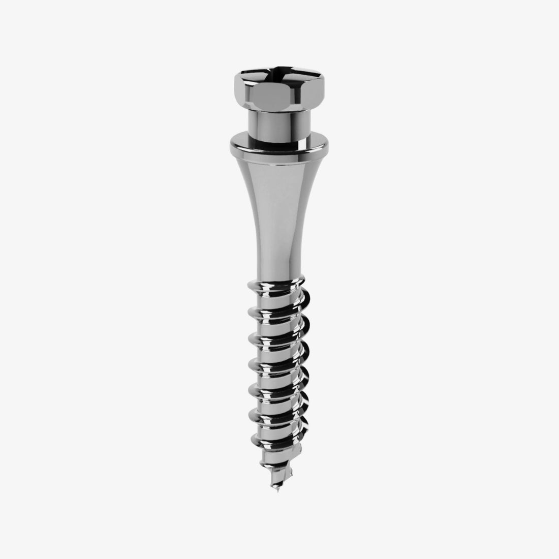
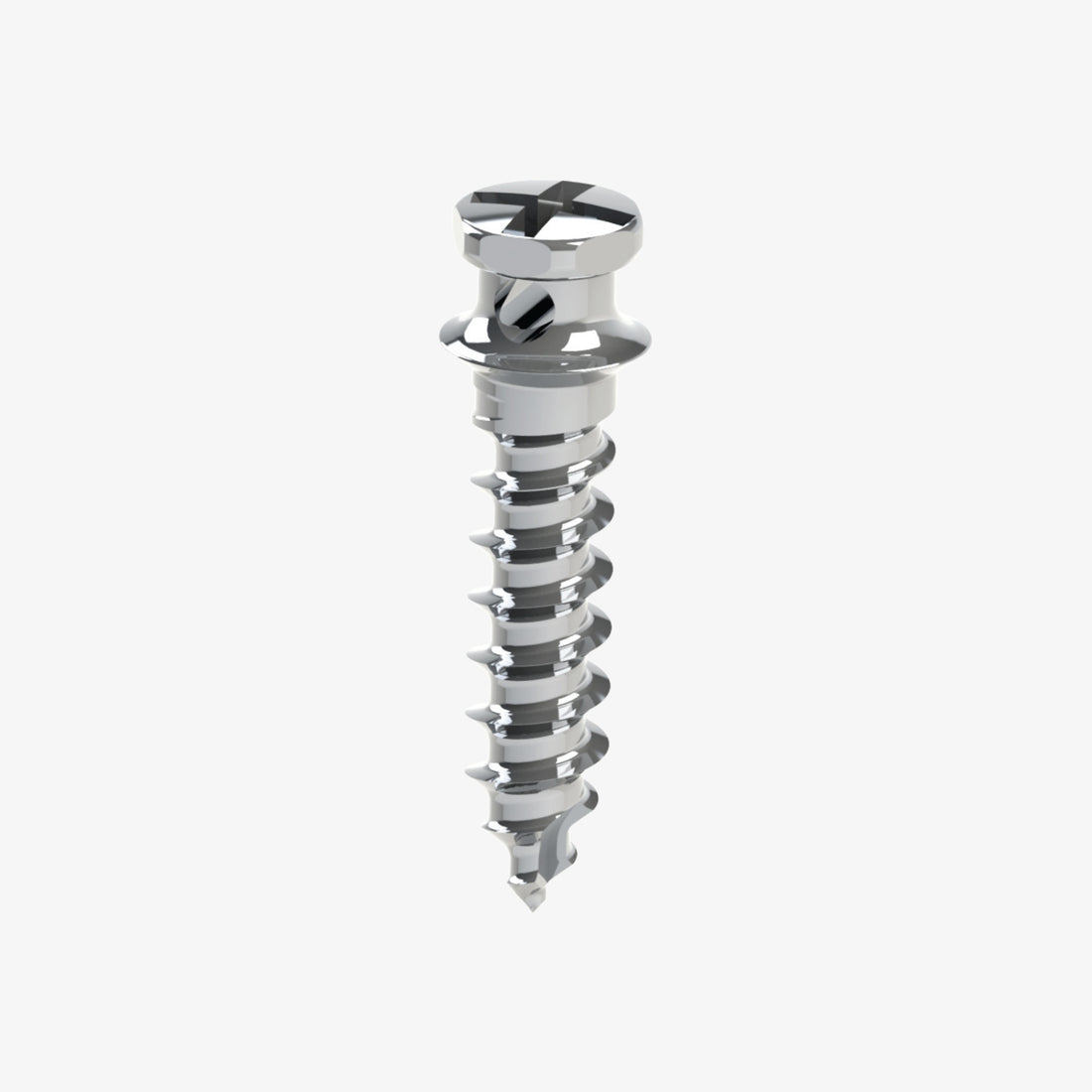
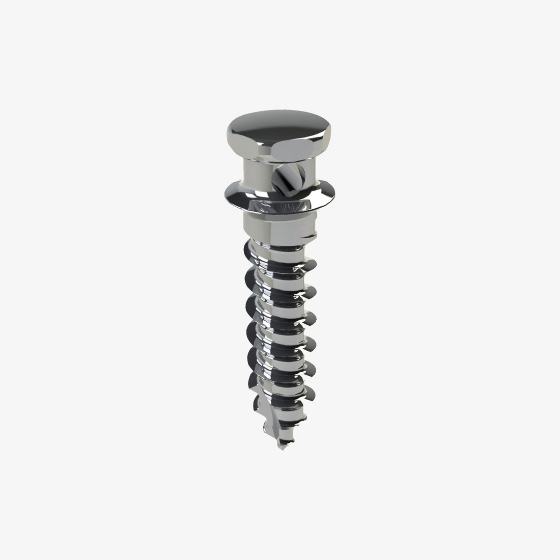
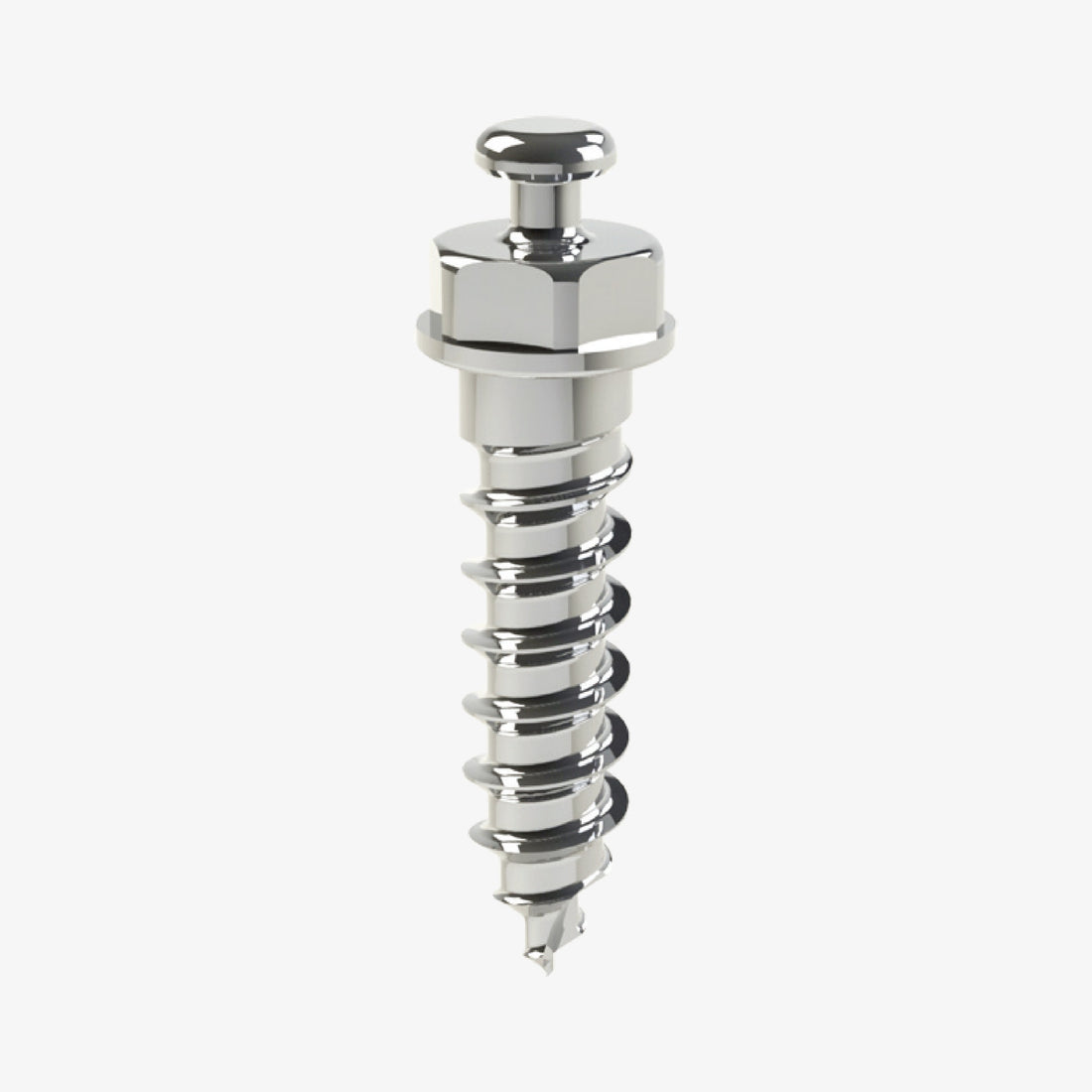
Clinical Example: Palatal Disjunction with JS Palatal Mini-Implants
A prime example of digital guidance enhancing precision is in the use of JS palatal mini-implants for skeletally-supported rapid palatal expansion (RPE), also known as MARPE (Mini-implant Assisted Rapid Palatal Expansion) or MSE (Maxillary Skeletal Expander). This technique is particularly effective in adolescent and adult patients where the midpalatal suture is already fused or significantly ossified, making traditional tooth-borne expanders less effective or leading to unwanted dental side effects.
Here's how digital guidance and laboratory collaboration streamline the process:
1. Initial Assessment and CBCT:
The orthodontist evaluates the patient's need for maxillary expansion, assessing the midpalatal suture maturity and bone thickness with a CBCT scan. This scan is crucial for identifying optimal mini-implant placement sites that avoid the nasal cavity, tooth roots, and offer sufficient bone volume.
2. Digital Planning (Orthodontist & Lab):
l The CBCT data is uploaded to 3D planning software.
l The orthodontist and the dental laboratory technician collaborate virtually. The software allows them to virtually place the JS palatal mini-implants (typically 2 or 4) in the thickest part of the palatal bone, perpendicular to the palate, ensuring maximum stability and avoiding vital structures.
l Simultaneously, the skeletally-borne expander device (e.g., a Hyrax-type expander) is virtually designed to connect directly to the heads of the planned mini-implants. This ensures a perfect fit and efficient transfer of expansive forces directly to the skeleton, bypassing the teeth.
l This virtual planning ensures that the mini-implants are positioned precisely to support the expander, allowing for predictable disjunction of the midpalatal suture.
3. Surgical Guide Fabrication: Based on the precise virtual planning, a 3D-printed surgical guide is fabricated. This guide, often tooth-borne or even mucosa-supported, incorporates sleeves that accurately dictate the drill path for the JS Palatal mini-implants, ensuring their exact position and angulation determined during planning.
4. Guided Mini-Implant Placement: The oral surgeon uses the custom surgical guide to place the JS Palatal mini-implant(s) with high precision. This minimizes the risk of root perforation or other complications and ensures the mini-implants are perfectly oriented for the subsequent attachment of the expander.
5. Laboratory Fabrication of Expander: Once the mini-implants are successfully placed (or even before placement, based on the virtual plan and printed models), the dental laboratory fabricates the custom expander. The expander is designed with connecting arms that precisely fit over the heads of the now-placed JS Palatal mini-implants, ensuring a passive and stable fit.
6. Expander Activation and Disjunction: After a short healing period, the expander is cemented (if tooth-borne) or directly connected to the mini-implants. The orthodontist then activates the expander, initiating controlled and predictable skeletal expansion by applying forces directly to the palate via the mini-implants.
This systematic, digitally guided approach, particularly with the JS Palatal mini-implant system, significantly enhances the success rate of skeletal expansion, reduces patient discomfort, and minimizes unwanted dental side effects, making it a gold standard for challenging disjunction cases.
Advantages of Digital Guided Skeletal Anchorage
The adoption of digital guided techniques offers a multitude of benefits for both clinicians and patients:
l Enhanced Precision and Accuracy: Drastically reduces the risk of misplacement and potential damage to adjacent structures.
l Increased Predictability: Ensures that the chosen anchorage system will perform as intended, leading to more predictable and efficient tooth movements.
l Improved Safety: Minimizes iatrogenic risks, enhancing patient safety during the procedure.
l Reduced Chair Time: While planning is more extensive, the actual surgical placement can often be faster and more straightforward.
l Optimal Biomechanics: Allows for the selection of anchorage sites that are ideally suited for the specific orthodontic forces required.
l Better Patient Communication: The 3D models and planning visuals can be used to educate patients about their treatment plan, improving understanding and compliance.
l Expanded Treatment Possibilities: Enables clinicians to confidently undertake more complex cases that might have been considered too risky with freehand techniques.
The Future is Guided
Digital guided skeletal anchorage represents a significant leap forward in orthodontic treatment. By seamlessly integrating diagnostic imaging with advanced planning and fabrication technologies, orthodontists can achieve a level of precision and predictability that was once unimaginable. As the technology continues to evolve, we can expect even greater refinements, further enhancing the safety, efficiency, and effectiveness of skeletal anchorage in orthodontics. The future of precise tooth movement is undoubtedly guided.


 0745 100 497


