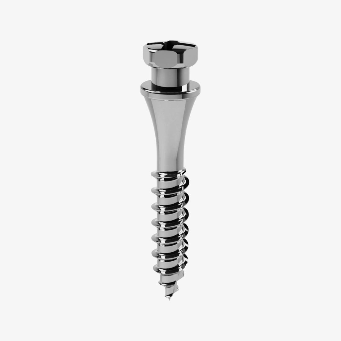
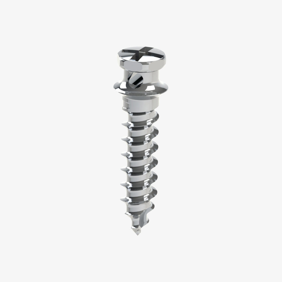
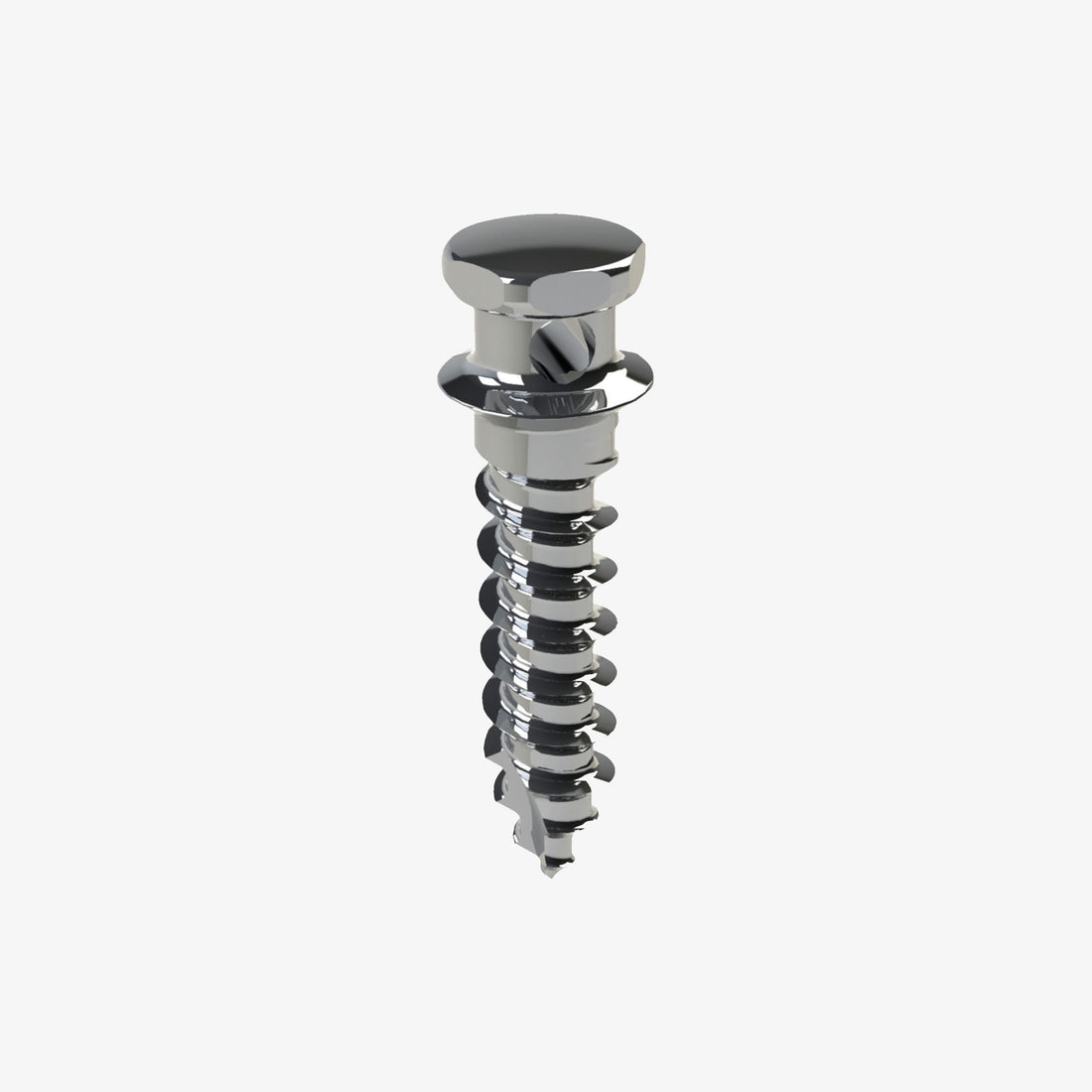
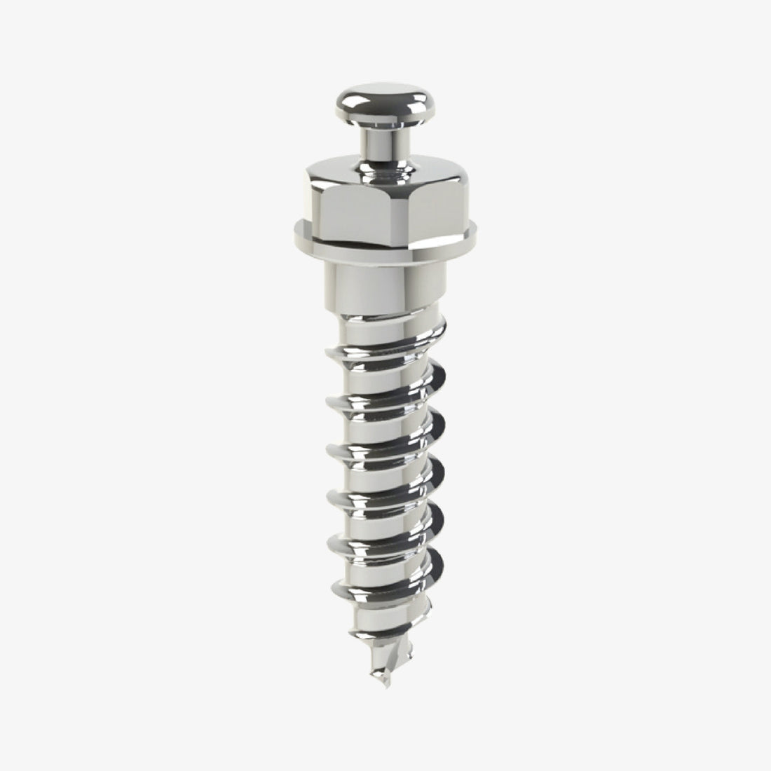
Mini implants, also known as Temporary Anchorage Devices (TADs), have revolutionized modern orthodontics by providing an efficient and stable method for applying corrective forces. These devices are particularly useful in complex cases where traditional braces might fall short. The placement of mini implants requires precise knowledge of various anatomical, mechanical, and biological factors to ensure optimal treatment outcomes. In this article, we will explore the factors that impact mini implant placement, including bone quality, mucosa considerations, root proximity, insertion techniques, and implant design.
I. Introduction to Mini Implants
Mini implants have become a fundamental tool in orthodontic treatment, offering significant advantages in both adult and pediatric care. Unlike traditional methods that rely on braces, TADs are used to provide anchorage for tooth movement without affecting the rest of the dental arch. These devices are placed into the bone, allowing the orthodontist to move teeth more effectively.
What Are Mini Implants?
Mini implants are small screws made of biocompatible materials like titanium, which are inserted into the jawbone or other regions of the mouth to serve as anchorage points for orthodontic forces. The main purpose of mini implants is to provide a stable point from which the orthodontist can apply force to move teeth, often in cases where traditional methods, such as braces or bands, are inadequate.
II. Factors Influencing Mini Implant Placement
A. Bone Quality and Quantity
One of the most critical factors in the success of mini implant placement is the quality and quantity of the bone in which the implant is inserted. The strength of the bone dictates how securely the implant will be anchored, affecting the stability and longevity of the mini implant.
Bone Quality:
-
D2 and D3 are considered ideal bone qualities for mini implant placement, offering a good balance between strength and flexibility. D2 is classified as dense bone, while D3 is slightly less dense but still suitable for stable anchorage.
Bone Quantity:
-
Imaging techniques such as CBCT (Cone Beam Computed Tomography) or CT scans allow clinicians to measure the quantity of bone available for mini implant insertion. The amount of bone directly influences implant length and positioning.
Source:
-
Al-Bitar, M., et al. (2017). "Bone Quality in Orthodontic Mini Implants: Influence on Anchorage and Stability." Journal of Orthodontic Science.
B. Mucosa Considerations
The type of mucosa in the area where mini implants are placed significantly affects the healing and success of the implant. There are two types of mucosa to consider:
-
Attached Mucosa: This type of mucosa is firmly connected to the underlying bone, making it ideal for mini implant placement due to its stability and reduced movement during orthodontic forces.
-
Mobile Mucosa: This type is not attached to the underlying bone, making it less stable and more prone to irritation and inflammation when subjected to the forces applied by mini implants.
Mini implants should always be placed in attached mucosa or at the boundary between attached and mobile mucosa to minimize complications. Additionally, frenulum proximity should be considered, as implants placed too close to the frenulum may irritate soft tissues and cause inflammation.
Source:
-
Kim, H., et al. (2014). "The Effect of Mucosal Attachment on Orthodontic Mini Implant Stability." Journal of Periodontology.
C. Root Proximity
Careful attention should be paid to the proximity of the mini implant to adjacent teeth, specifically their roots. The ideal implant location is one that ensures at least 1mm of bone surrounding the implant and at least 0.5mm between the implant and the root of adjacent teeth.
Techniques for Avoiding Root Damage:
-
Clinicians use periodontal probes to palpate the surface of the roots, which helps determine the exact space available for the mini implant.
-
Radiographic imaging allows for a detailed view of root positioning relative to the potential implant site, reducing the risk of root damage.
Source:
-
Tuna, M., et al. (2012). "Periodontal Probing and Root Spacing in Mini Implant Placement." American Journal of Orthodontics.
III. Surgical Techniques for Mini Implant Insertion
A. Implant Placement Methods
The successful placement of mini implants requires precise technique to ensure both stability and long-term success. The basic steps of insertion include:
-
Preparation:
-
Thorough assessment using imaging techniques (such as CBCT scans) to evaluate bone quality, root proximity, and mucosal type.
-
Incision and Site Preparation:
-
In some cases, a small incision is made to expose the bone, while in others, the implant can be placed through the mucosa directly.
-
Angle of Insertion:
-
The angle of insertion is crucial for achieving optimal stability in the cortical bone. Generally, a 45-degree angle is ideal, with adjustments made depending on whether the implant is placed in the maxilla or mandible.
-
Implant Insertion:
-
The implant is then placed, ensuring it remains perpendicular to the bone for initial stability. Adjustments are made based on bone and mucosa thickness.
Source:
-
Gambarini, G., et al. (2015). "Techniques for the Insertion of Mini Implants in Orthodontics." Orthodontics and Craniofacial Research.
B. Post-Insertion Care
Once the mini implants are placed, proper post-operative care is essential for promoting healing and avoiding complications such as infection or implant failure.
-
Soft Tissue Care: Keep the surrounding tissues clean and free from irritation to avoid inflammation.
-
Bone Healing: Patients may need to limit physical activity to ensure proper osseointegration (bone healing around the implant).
IV. Adjacent Anatomical Structures and Precautions
While planning mini implant placement, it's essential to be mindful of surrounding structures, including nerves, sinuses, and fossa. For example, in the palate, the palatal nerve must be avoided, and in the mandible, the mental nerve and inferior alveolar nerve are significant concerns.
One effective region for mini implant placement is the T-zone in the palate, as it is a stable location away from these critical structures.
Source:
-
Vas, R., Ludburn, R. (2013). "Safe Zones for Implant Placement in Orthodontics." International Journal of Oral and Maxillofacial Surgery.
V. Choosing the Right Implant
After considering the bone quality, mucosa, root spacing, and surrounding structures, it’s important to select the appropriate implant diameter, length, and design to match the treatment goals. Larger implants may be required for cases with insufficient bone, while smaller ones are suitable for less invasive cases.
VI. Conclusion
Mini implants offer orthodontists a valuable tool for enhancing tooth movement and treatment outcomes. Successful implant placement requires an understanding of bone quality, mucosa type, root proximity, and anatomical structures. By considering these factors and using advanced imaging and techniques, orthodontists can ensure the long-term success and stability of mini implants.
References
-
Al-Bitar, M., et al. (2017). "Bone Quality in Orthodontic Mini Implants: Influence on Anchorage and Stability." Journal of Orthodontic Science.
-
Kim, H., et al. (2014). "The Effect of Mucosal Attachment on Orthodontic Mini Implant Stability." Journal of Periodontology.
-
Tuna, M., et al. (2012). "Periodontal Probing and Root Spacing in Mini Implant Placement." American Journal of Orthodontics.
-
Gambarini, G., et al. (2015). "Techniques for the Insertion of Mini Implants in Orthodontics." Orthodontics and Craniofacial Research.
-
Vas, R., Ludburn, R. (2013). "Safe Zones for Implant Placement in Orthodontics." International Journal of Oral and Maxillofacial Surgery.
-
D’souza, R., et al. (2020). "Evaluation of Bone Density and Its Impact on Mini Implant Success in Orthodontics." Orthodontic Journal of India.


 0745 100 497


